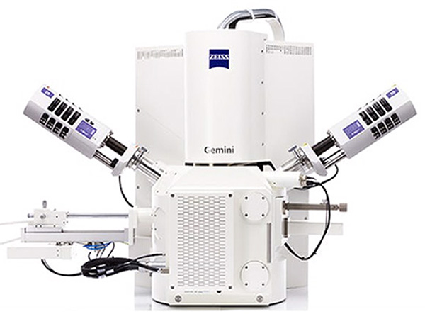
ZEISS SIGMA
Field Emission Scanning Electron Microscope
Overview
Since the introduction of the ZEISS GEMINI column design in 1994, ZEISS continues to set the standard for Low keV performance and ease of use, The ZEISS SIGMA Field Emission Electron Microscope provides the best price to performance ratio in the industry. The ZEISS Sigma family combines field emission SEM (FE-SEM) technology with an excellent user experience. The SIGMA enables users with SIGMA’s intuitive 4-step workflow structuring imaging and analysis routines, increasing productivity. End users will capture more data, faster than ever before. Choose from a variety of detector options to tailor SIGMA precisely to your applications: you can image particles, surfaces, nanostructures, thin films, coatings and layers. With the Sigma family you enter the world of high-end imaging.
Unique Features
Excellence in price and performance
- Best-in-class EDS geometry delivering superb analytical performance
- Superior Low keV performance
- Best in class variable pressure imaging and performance
- Excellent sample handling capability
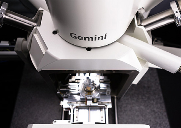
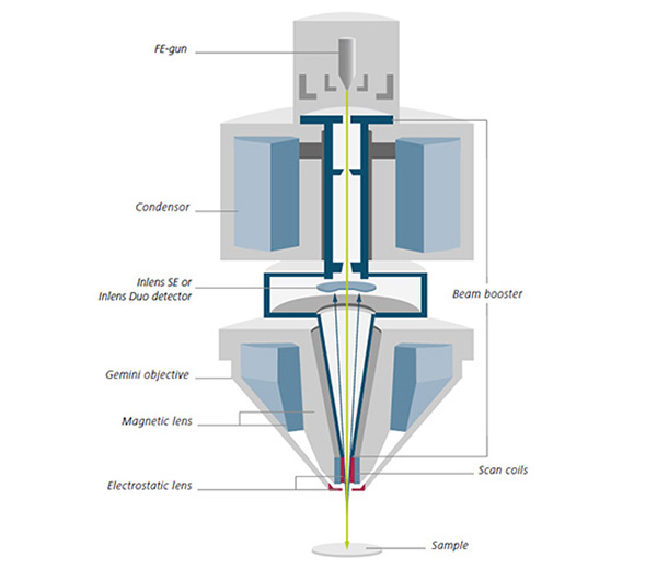
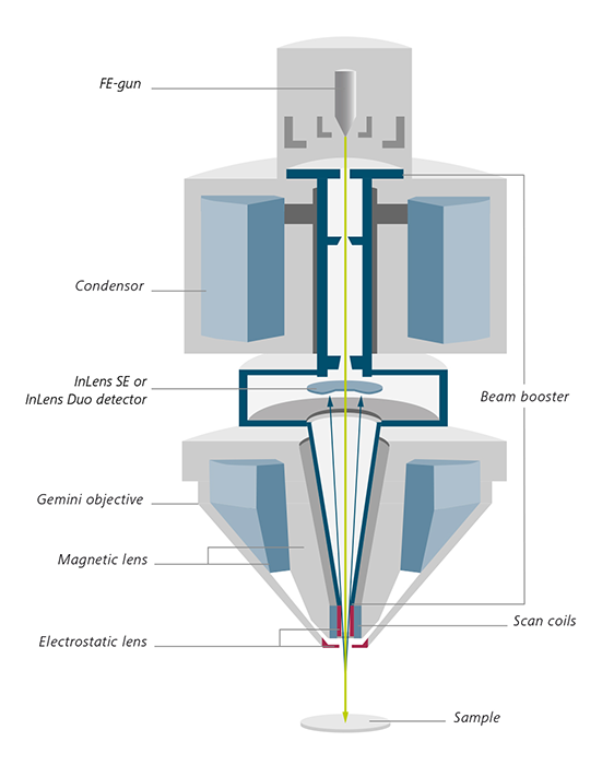
Your Insight into the Technology Behind It
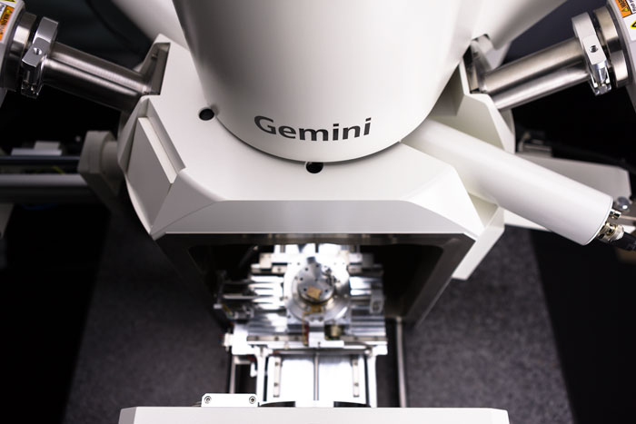
Based on Proven Gemini Technology
The Sigma family is based on more than 20 years of perfecting Gemini technology. You can count on complete and efficient detection, excellent resolution and unsurpassed ease-of-use. The Gemini objective lens design combines electrostatic and magnetic fields to maximize optical performance while reducing field influences at the sample to a minimum. This enables excellent imaging, even on challenging samples such as magnetic materials. The Gemini detection concept ensures efficient signal detection by detecting secondary (SE) and/or backscattered (BSE) electrons. This so-called lnlens detector is arranged on the optical axis, which reduces the need for realignment and thus minimizes time-to image. Gemini beam booster technology guarantees small probe sizes and high signal-to-noise ratios, right down to ultra-low accelerating voltages. It also minimizes system sensitivity to external stray fields by keeping the beam at high voltage throughout the column until its final deceleration.
Highlights
Flexible Detection for Clear Images
- Tailor Sigma to your needs using the latest detection technology and characterize all of your samples
- Acquire topographical and compositional informauon with the optional Inlens Duo detector.
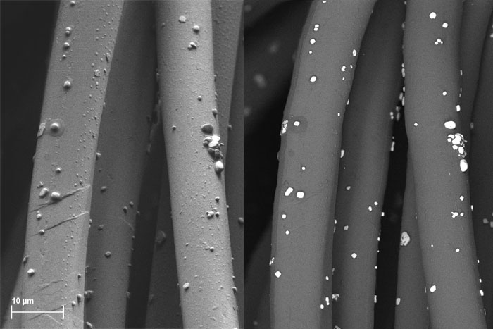
- Enjoy a new generation of secondary electron (SE) detectors. Obtain images with up to 50% more signal. Benefit from the novel C2D and VPSE detectors of Sigma in variable pressure mode: working at low vacuum, you can expect crisp images with up to 85% more contrast.
Automate and Speed up Your Workflow
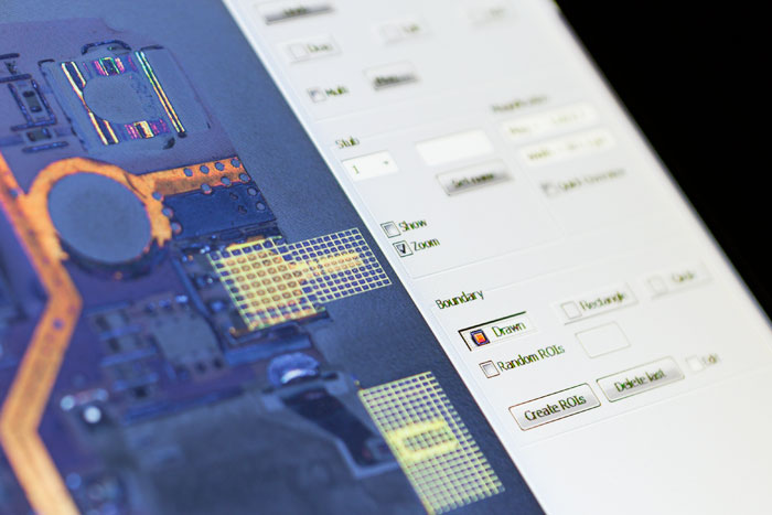
- A 4-step workflow lets you control all the functionality of your Sigma. Benefit from fast time-to-image and save time on training - especially in a multi-user environment.
- First, navigate your sample and then set optimal imaging conditions
- Next. automatically acquire images across multiple samples utilizing regions of interest (ROIs). Finally use the workflow's last step for contextual visualization of your results.
Perform Advanced Analytical Microscopy
- Combine scanning electron microscopy and elemental analytics: the best-in-class EDS geometry of Sigma increases your analytical productivity, especially on beam sensitive samples.
- Get analytical data at half the probe current and twice the speed.
- Achieve complete, shadow·free analytics in your FE-SEM. Profit from using a short analytical working distance of 8.5 mm and a take-off angle of 35°
Accessories
SmartEDX
Discover Embedded Energy Dispersive X-ray Spectroscopy Analysis

- Optimization for routine microanalysis applications and detection of low energy X-rays from light elements thanks to superior transmissivity of the silicon nitride window
- Workflow-guided graphical user interface improves ease-of-use and repeatability in multi-user environments
- Total service and system support by a ZEISS engineer is giving you a one-stop shop for installation, preventive maintenance and warranty
Raman Imaging and Scanning Electron Microscopy
Reap the Benefits of Fully Integrated RISE
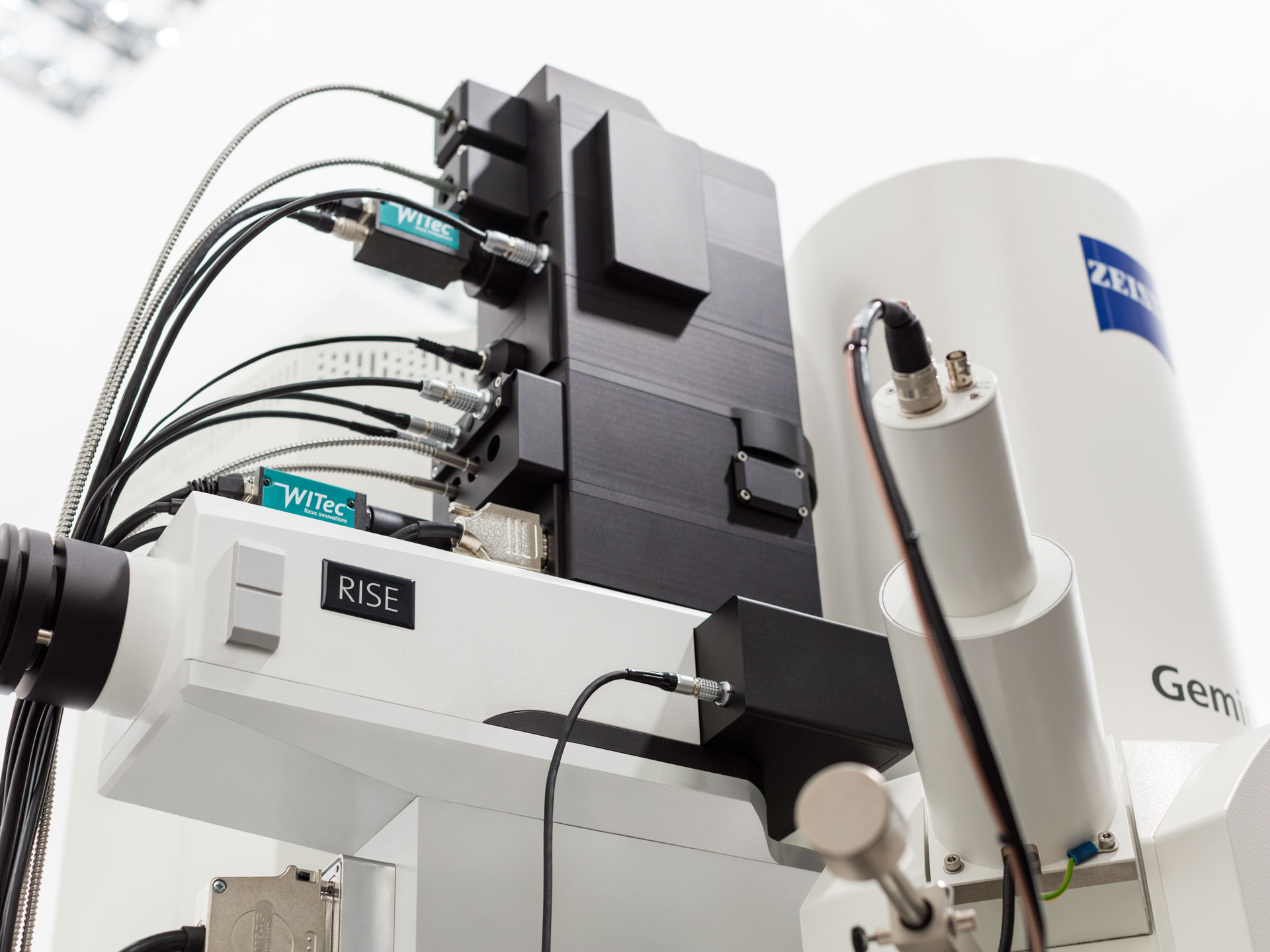
- Recognize molecular and crystallographic information
- Perform 3D analyis and correlate SEM imaging, with Raman mapping and EDS data if appropriate
- Fully integrated RISE lets you take advantage of both best-in-class SEM and Raman systems
Software
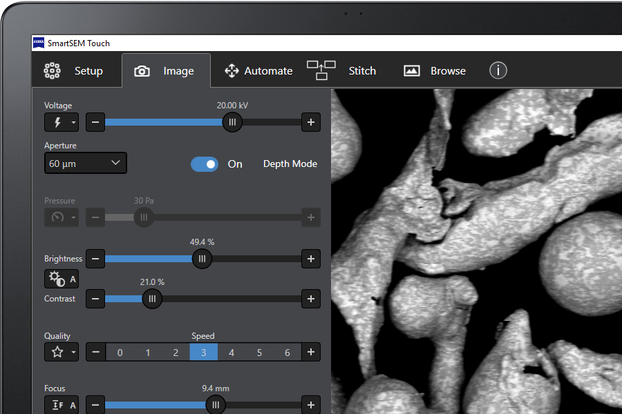
ZEISS SmartSEM Touch - Get more Hands on Deck
SmartSEM Touch, an add-on to the established operation system, is a simplified user-interface for multi-user environments. It comes wtth easy operation for both expenenced and novice users. Depending on the actual laboratory environment, operation of the SEM can be the exclusive domain of expert electron microscopists. But this situation is challenged by the very common necessity that non-expert users, such as students, trainees. or quality engineers, also require data from the SEM. Sigma 300 and Sigma 300 VP take both requirements into account, with user interface options that cater to the operational needs of experienced microscopists as well as non-microscopists.
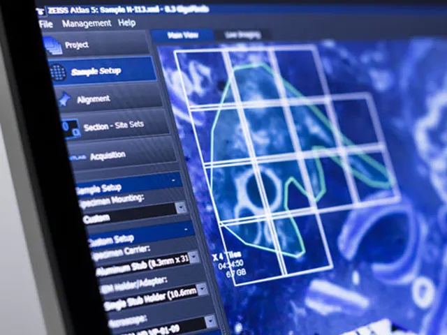
ZEISS Atlas 5 - Master Your Multi-scale Challenge
Atlas 5 makes your life easier: create comprehensive multi-scale, multi-modal images with a sample-centric correlative environment. Atlas 5 is the powerful yet intuitive hardware and software package that extends the capacity of your scanning electron microscope.
Visualization and Analysis Software
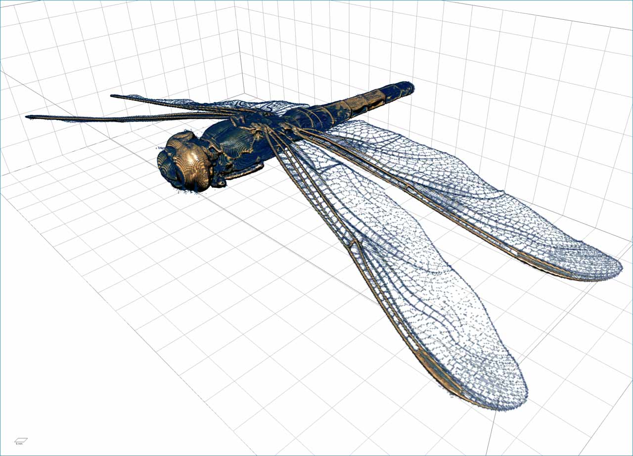
ZEISS recommends Dragonfly Pro from Object Research Systems (ORS)An advanced analysis and visualization software solution for your 3D data acquired by a variety of technologies includ1ng X-ray, FIB-SEM, SEM and helium ion microscopy.
Formerly Visual SI Advanced, Dragonfly Pro delivers high-definition visualization techniques and industry-leading graphics. Dragonfly Pro supports customization through easy to use Python scripting. Users now have total control of their 3D data post-processing environment and workflows.