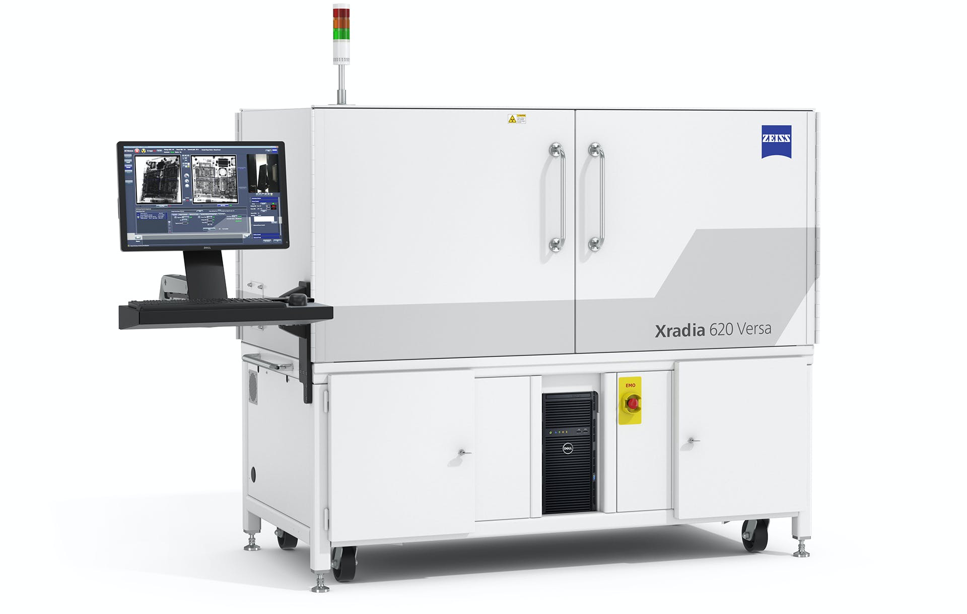
ZEISS Xradia 515, 615 and 730 Versa 3D X-ray Microscopy for Faster Sub-Micron Imaging of Intact Samples
Overview
Move beyond exploration to achieve your moments of discovery unlocking new degrees of versatility for your scientific and industrial research with the most advanced 3D X-ray microscope models in the ZEISS Xradia Versa family. Building on industry-best resolution and contrast, the ZEISS Versa expands the boundaries of your non-destructive sub-micron scale imaging
- Higher flux and faster scans without compromising resolution
- True spatial resolution to 450 nm with a minimum achievable voxel size of 40 nm
- High resolution across a broad range of sample types, sizes, and working distances
- In situ imaging for non-destructive characterization of microstructures in controlled environments and over time
- Upgradeable and extendible with future innovations and developments
- Throughput with image quality
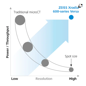
Highest Resolution without Compromise
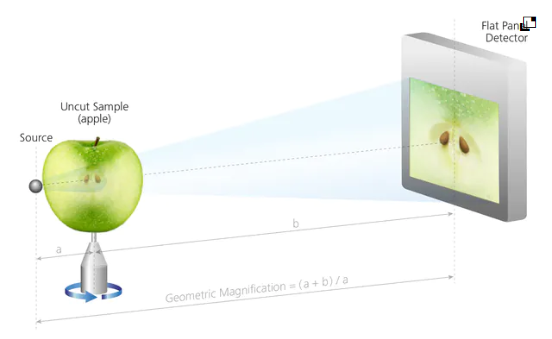
Standard X-ray computed tomography (CT) is limited to small sample sizes when imaging at high resolution; due to the geometric nature of magnification. Maintaining high resolution for larger samples is impossible due to the longer working distances required. High resolution imaging in CT systems also requires low X-ray flux, reducing the throughput of the measurement. This limits the practical application of maximum resolution claimed by most CT manufacturers.
The ZEISS Versa overcomes these trade-offs by integrating dual-stage magnification architecture with high flux X-ray source technology.
ZEISS specifies true spatial resolution, bringing a standard measure of microscope performance to 3D X-ray measurement. Spatial resolution refers to the minimum separation at which two features can be resolved by an imaging system.The ZEISS Series Versa systems obtain true spatial resolution down to 450 nm with a minimum achievable voxel size of 40 nm.
ZEISS X-ray Microscopes
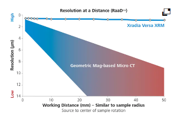
The Versatile Advantage of RaaD
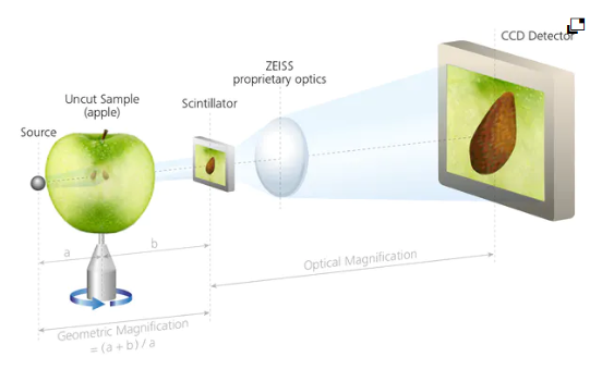
ZEISS Xradia Versa uses a two-stage magnification architecture to enable sub-micron resolution imaging at large working distances (Resolution at a Distance) for a diverse set of sample sizes and types. Images are initially magnified via geometric projection as with conventional microCT, the projected image is cast onto a scintillator, converting X-rays to a visible light image which is then optically magnified by microscope optics before acquisition by a CCD detector.
With more X-ray photons available, the ZEISS Versa provides faster time to results for the widest range of sample sizes and types, without compromising resolution.
Conventional microCT Architecture
ZEISS XRM Two-stage Magnification Architecture
Accessories
Extend the Range of Possibility for Advanced
Material Characterization in 3D
LabDCT
Unlocking Crystallographic Information
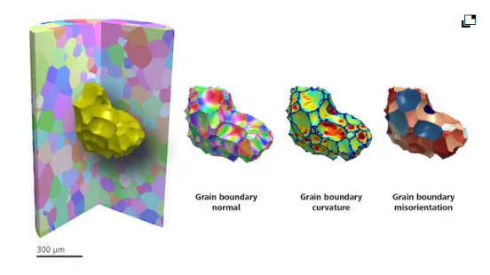
LabDCT (Diffraction Contrast Tomography), available exclusively on Xradia 620 Versa, enables non-destructive mapping of orientation and microstructure in 3D. Direct visualization of 3D crystallographic grain orientation opens up a new dimension in the characterization of polycrystalline materials like metal alloys, geomaterials, ceramics, or pharmaceuticals.
- LabDCT now supports specimens with crystal structures from high cubic symmetry to systems with lower symmetry such as monoclinic materials.
- Obtain comprehensive 3D microstructure analysis from large volumes down to local individual grain boundary analysis.
- Investigate microstructural evolution with 4D imaging experiments.
- Combine 3D crystallographic information with 3D microstructural features.
- Combine modalities to understand structure-property relationships.
Flat Panel Extension
Image even larger samples with high throughput
Optional Flat Panel Extension (FPX) delivers large-sample, high throughput scanning with ZEISS best-in-class image quality. FPX enhances imaging flexibility and creates workflow efficiencies with an all-in-one system for industrial and academic research.
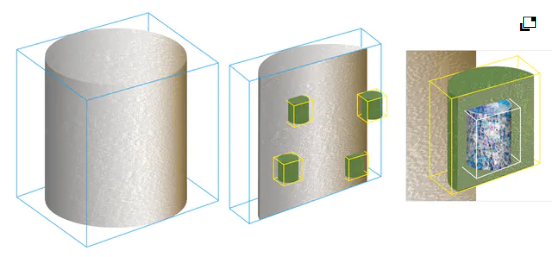
Scout-and-Zoom is a unique capability of ZEISS X-ray microscopes that leverages FPX to perform exploratory “Scout” scans across a large field of view to identify interior regions of interest for higher resolution “Zoom” scans without complex sample preparation.
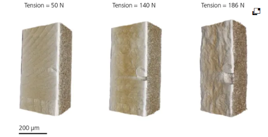
In Situ experiments
Push the limits for scientific advancement
ZEISS Xradia Versa provides the industry’s premier 3D imaging solution for the widest variety of in situ rigs, from high pressure flow cells to tension, compression and thermal stages.
To accommodate various types of in situ apparatus, such experiments require samples to be mounted further away from the X-ray source. With traditional microCT systems, this significantly limits the resolution achievable during such measurements. ZEISS X-ray microscopes are uniquely capable of Resolution at a Distance (RaaD) technology, which give the highest fidelity of 3D structural information during in situ imaging.